Biophysical Methods
How the key biophysical techniques work — from label-free biosensors that measure real-time kinetics, to calorimetry, in-solution binding assays, and structural characterization methods.
Label-Free Kinetics
Surface-based biosensors that measure real-time binding kinetics (kon, koff, KD) without labelling the target.
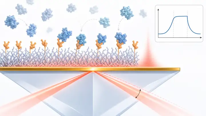
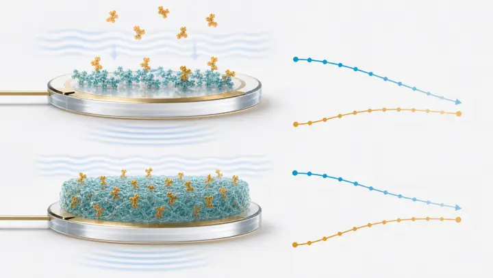
Surface Plasmon Resonance (SPR)
Surface plasmons, evanescent waves, and how gold films detect molecular binding in real-time.
Bio-Layer Interferometry (BLI)
White light interference on fiber-optic biosensors. Dip-and-read format for high throughput.
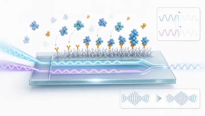
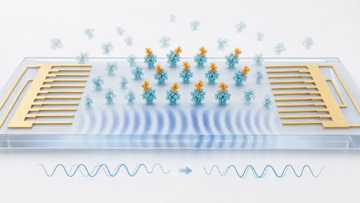
Grating-Coupled Interferometry (GCI)
Planar waveguide biosensor using an evanescent field over a longer interaction path than SPR, with interferometric phase-shift readout for high sensitivity.
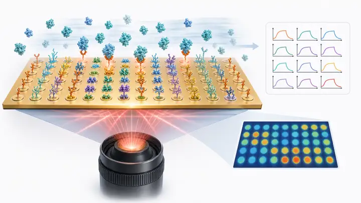
SPR Imaging (SPRi)
Array-based SPR: image hundreds of spots simultaneously. Parallel kinetics, spatial referencing, and sensorgram maps on a single chip.
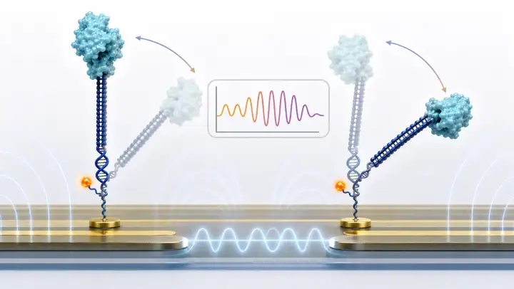
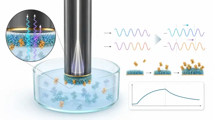
switchSENSE — heliX+
Label-free surface kinetics on electrically actuated DNA nanolevers. The reporter dye sits on the DNA adapter strand (not the target), so kon, koff, and KD are measured without labelling the protein — and conformational changes upon binding are accessible as a bonus.
Focal Molography
Coherent diffraction from a molecular grating. Self-referencing readout strongly suppresses bulk RI drift and uncorrelated non-specific adsorption.
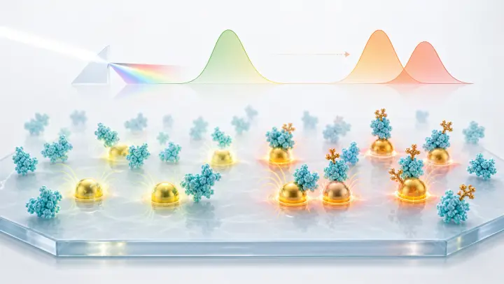
Localized Surface Plasmon Resonance (LSPR)
Gold nanoparticles absorb light at a shape-dependent wavelength. The physics behind home pregnancy tests, rapid diagnostics, and ultrasensitive near-field biosensors.
On-Cell Kinetics
Measure real-time binding kinetics directly on native cell-surface receptors — capturing avidity and cell-to-cell heterogeneity that purified-protein assays miss.
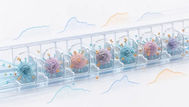
heliXcyto — Single-Cell Interaction Cytometry
Trap individual cells in polymer cages and measure real-time kon, koff, and KD on native cell-surface receptors. Captures avidity and cell-to-cell heterogeneity that purified-protein biosensors miss.

LigandTracer — Real-Time Binding on Adherent Cells
Ridgeview Instruments rotates a tilted petri dish under a fluorescent or radioactive tracer detector. Hours-to-days kinetics on live adherent cells — ideal for slow off-rates, radioligands, and native receptor pharmacology.
Thermodynamic Methods
Measure binding energetics — enthalpy, entropy, heat capacity, and thermal stability.
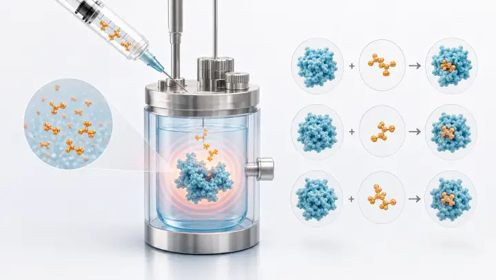
Isothermal Titration Calorimetry (ITC)
The gold standard for binding thermodynamics. Measure ΔH, ΔS, KD, and stoichiometry in a single experiment.
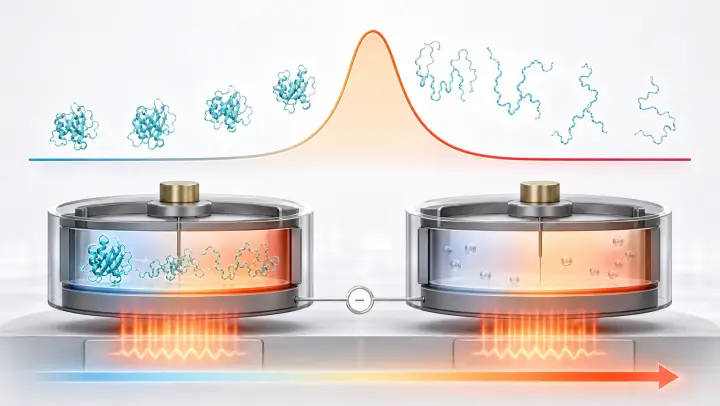
Differential Scanning Calorimetry (DSC)
Direct measurement of unfolding thermodynamics: Tm, ΔH, ΔCp, and the full stability curve ΔG(T). Gold standard for thermal characterization.
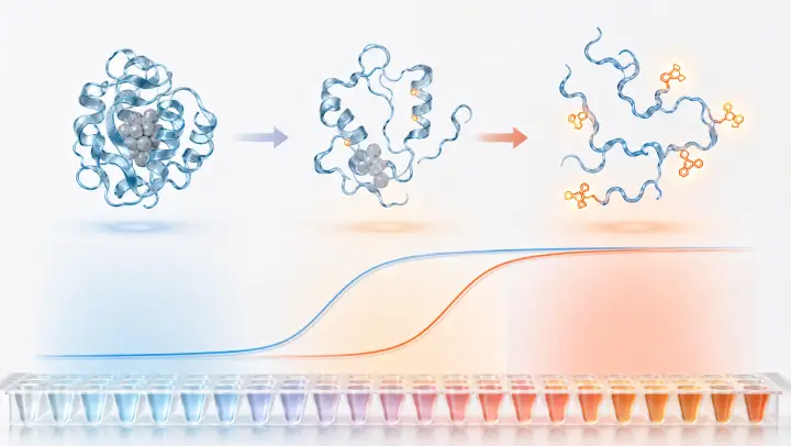
Thermal Shift Assay (TSA/DSF)
Protein stability screening with fluorescent dyes and intrinsic fluorescence. qPCR-based thermal melts, ΔTm for ligand binding, and nano-DSF.
In-Solution Binding Assays
Measure binding affinity and interactions in free solution — no surface immobilization required.
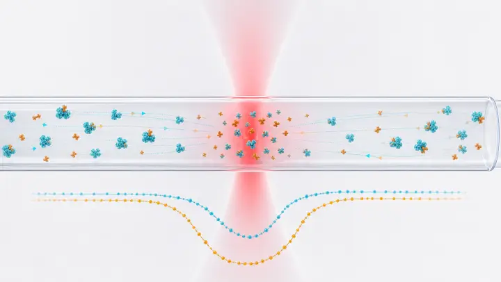
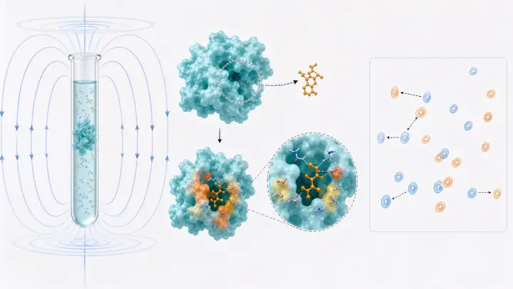
Microscale Thermophoresis (MST)
Measure binding affinity in free solution via thermophoresis. Requires only microliters of sample — ideal for targets that resist immobilization.
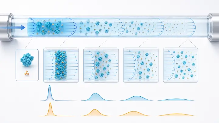
Flow-Induced Dispersion Analysis (FIDA)
Measure hydrodynamic radius and binding affinity from Taylor dispersion in a capillary. Works in crude matrices — serum, lysate, formulation buffer.
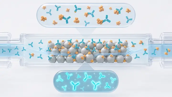
Kinetic Exclusion Assay (KinExA)
Solution-equilibrium K_D of unmodified binding pairs. Captures only free antibody from a pre-equilibrated mixture — pushes affinity measurement into the femtomolar range, well below SPR/BLI floors.
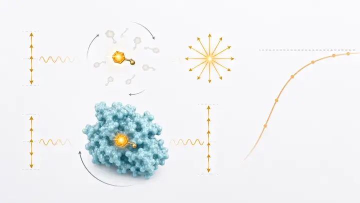
Fluorescence Polarization (FP/FA)
Molecular rotation meets binding detection. Polarized light, the Perrin equation, and high-throughput competition assays for drug discovery.
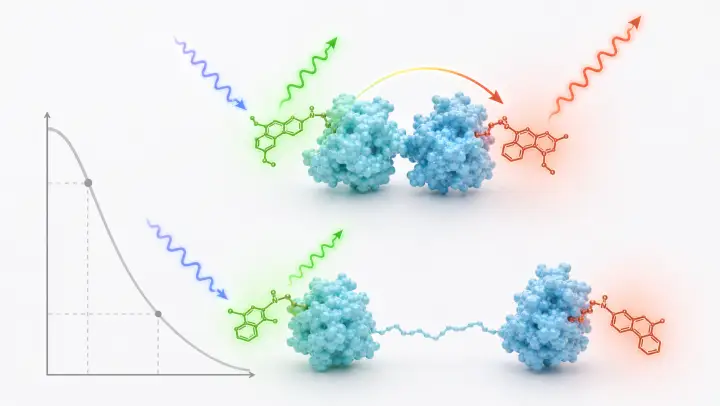
Förster Resonance Energy Transfer (FRET)
Distance-dependent energy transfer between fluorophores. TR-FRET for drug screening, smFRET for conformational dynamics.
Acoustic Biosensors
Measure mass and viscoelastic properties via piezoelectric resonance or surface waves.
Structural & Biophysical Characterization
Complementary methods for conformation, size, stoichiometry, and binding-site mapping.
NMR Spectroscopy
Atomic-resolution binding site mapping in solution. CSP titrations, STD screening, relaxation dispersion kinetics — no immobilization required.
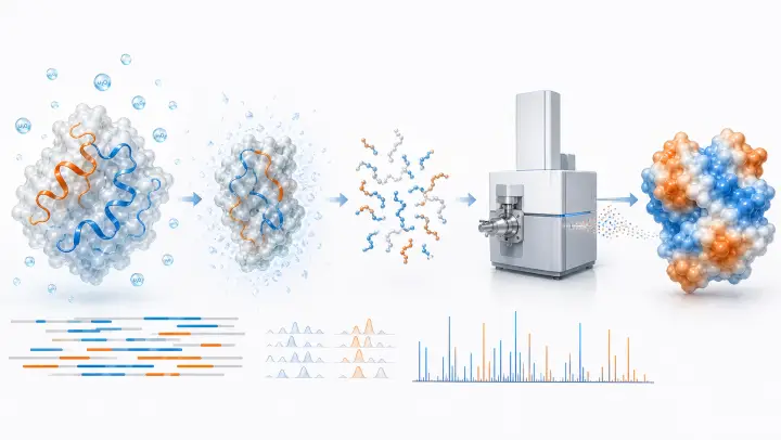
Hydrogen-Deuterium Exchange Mass Spectrometry (HDX-MS)
Map protein conformational dynamics by tracking backbone amide exchange with deuterium. Peptide-level resolution for epitope mapping, allostery, and biosimilar comparability.
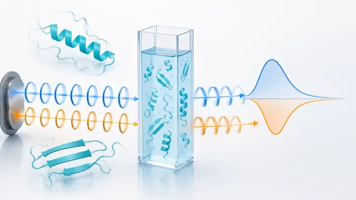
Circular Dichroism (CD)
Protein secondary structure from differential absorption of circularly polarized light. Thermal stability, conformational changes, and quality control — all in solution.
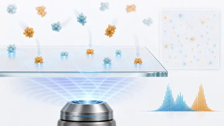
Mass Photometry
Single-molecule mass measurement by interferometric scattering. Weigh individual biomolecules as they land on glass — minutes of acquisition, nanomolar working concentrations.
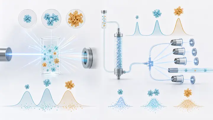
DLS & SEC-MALS
Hydrodynamic size, polydispersity, and absolute molecular weight. Quality control for every binding experiment.
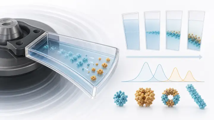
Analytical Ultracentrifugation (AUC)
Solution-state sedimentation analysis for c(s) distributions, buoyant molar mass, oligomerization, and hydrodynamic heterogeneity.
Cross-Method Resources
Concepts and comparisons that span multiple techniques.
The Sensorgram
Physics meets biology. Interactive simulator of baseline, association, dissociation, and artifacts — the shared language of SPR, BLI, and GCI.
Method Comparison
Side-by-side comparison of SPR, BLI, GCI, ITC, and MST — sensitivity, throughput, kinetics, and best use cases.
Continue Learning
Now that you understand the methods, explore the mathematical models used to fit binding data.